⚙
Automated TMA construction
Choose the right level of automation for your laboratory: semi-automatic, computer-assisted, computer-driven or fully automated.
ISENET develops Galileo Tissue Microarray instruments for biomarker validation, spatial omics, multiplexing and digital pathology workflows — from semi-automatic systems to fully automated high-throughput platforms.
Build precise TMAs while preserving the chain of information from donor block to TMA spot, slide image and downstream analysis.
Choose the right level of automation for your laboratory: semi-automatic, computer-assisted, computer-driven or fully automated.
Connect donor block ID, ROI, core coordinates, recipient position and analysis output using digital reports and export files.
Support biomarker studies, multiplex staining, spatial transcriptomics and digital pathology workflows using reproducible TMA layouts.
Each system is positioned around a clear laboratory need, making the product choice faster and more intuitive.
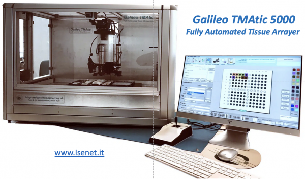
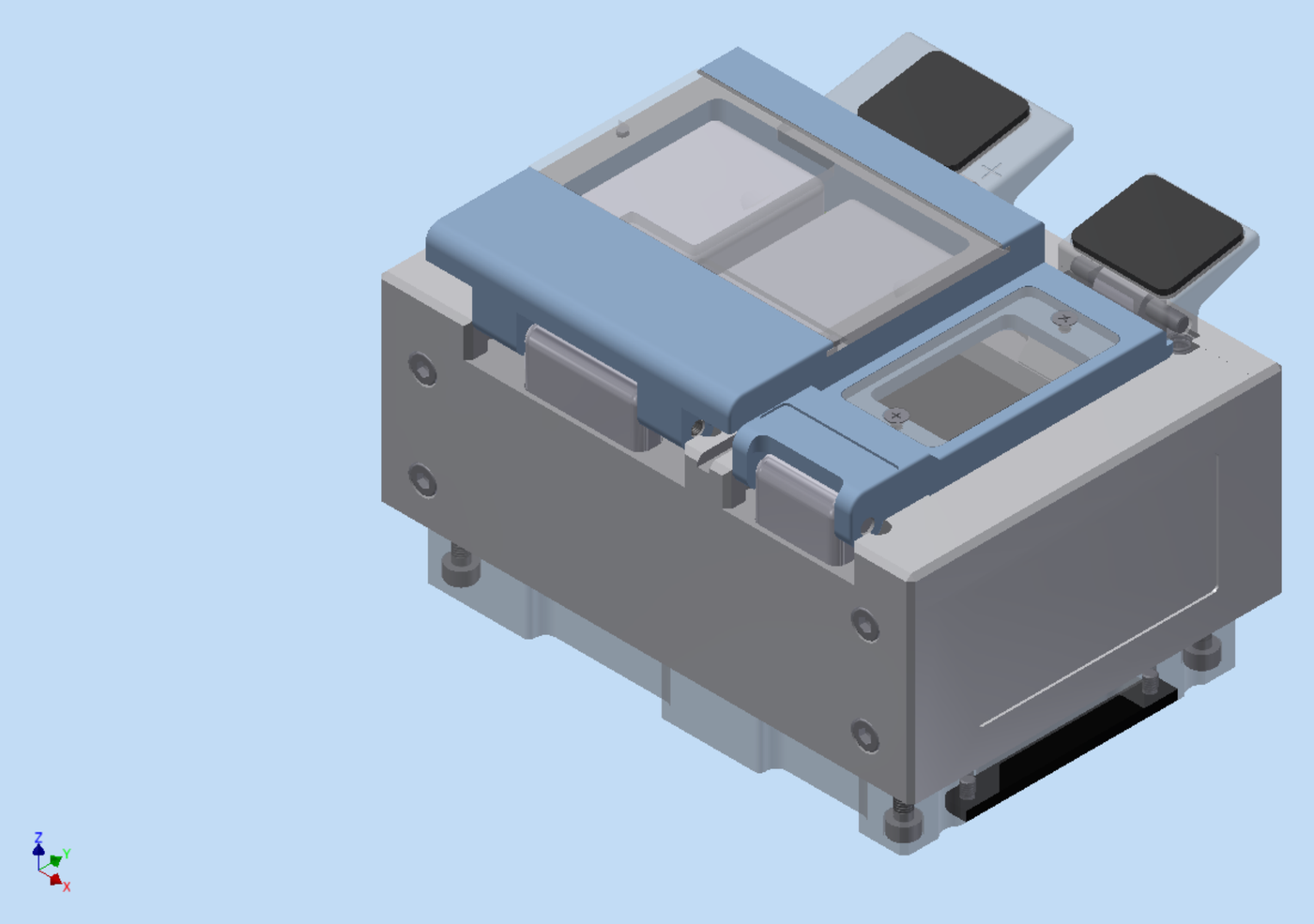
Fully automated TMA platform
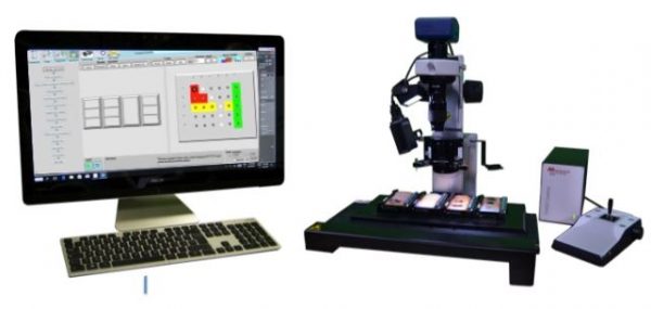
Computer-driven TMA system
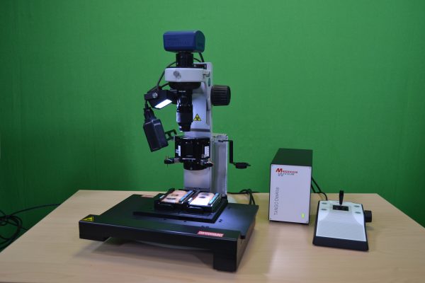
Computer-assisted TMA system
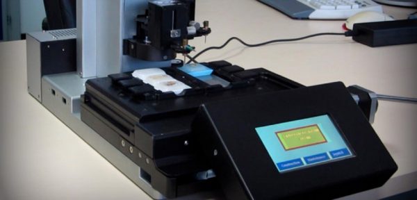
Semi-automatic TMA system
Accessory for frozen TMA construction
Galileo platforms support classic pathology research and the newest high-value applications in spatial biology.
Screen large cohorts with consistent tissue sampling and controlled TMA geometries.
Reduce per-sample assay costs by arranging multiple tissue samples on a single high-value slide.
Enable standardized comparative staining and image analysis across cases, controls and replicates.
A modern TMA workflow must preserve the link between the original donor tissue and the final digital result.
Acquire donor block or slide images for ROI selection.
Define tissue regions to sample and associate them with donor IDs.
Construct the recipient block using the selected Galileo platform.
Digitize TMA sections using compatible whole-slide scanners.
Use array maps and export files to support de-arraying and image analysis.
Instrument images and YouTube videos are taken from the existing ISENET web pages and ISENET brochure links, so your web developer can reuse the same WordPress Media Library assets.
Fully automated TMA platform.
Computer-driven TMA system.
Computer-assisted TMA system.
Semi-automatic TMA system.
Frozen TMA Module for compatible CK3600 / CK4600 systems.
Request a demo or ask ISENET for a product comparison based on your throughput, core size and digital pathology requirements.